Reported in the literature have a common pathogenesis of systemic sclerosis and cancer rate was 3-7%. Malignancies occurred after systemic sclerosis, scleroderma can occur before or simultaneously occur. In addition to lung cancer rarely found outside early systemic sclerosis, tumors in other organs seem to have these three cases. Studies have 2141 cases of systemic sclerosis patients, 66 cases of systemic sclerosis associated with 72 kinds of cancer, including 55 cases (85.3%) occurred after systemic sclerosis, only 17 cases (25.8%) prior to the occurrence of systemic sclerosis .
Systemic sclerosis associated with malignant lung cancer, breast cancer, lymphoma and leukemia the most common, and the incidence of lung cancer is elevated. Most lung cancer occurs in systemic sclerosis 10 years later, and most of lymphoma, leukemia, breast cancer occurred in the three years after systemic sclerosis. And its most common histologic type of adenocarcinoma.
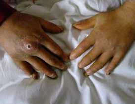
Systemic sclerosis is generally considered chronic organ fibrosis, especially prevalent in skin tumors, lung and gastrointestinal tract. Because of the sensitive site of the lesion has a wide range of immune complex deposition, it may also be related to an abnormal immune response, in the absence of immune surveillance, so that structural abnormalities of the lesion removal of carcinogens impaired, increased epithelial cell proliferation of malignant transformation under a variety of possible mechanisms, etc., so that cancer incidence. Also patients receiving immunosuppressive therapy may increase the risk of malignancy.
Furthermore, the formation of self-antigens of tumor cells, to induce autoantibodies in a particular stage of the disease, and the substrate can cause denaturation of collagen-induced systemic sclerosis or systemic sclerosis like symptoms.
Therefore, female patients in the first five years after scleroderma should be a breast cancer and early chest X-ray examination every year and should continue. Among those with pulmonary fibrosis, five years after the first chest X-ray examination should be carried out annually. To have reflux esophagitis, obstruction of the distal esophagus in patients with endoscopy and biopsy should be drawn. Patients who have received immunosuppressive therapy, blood and medicine should be a long-term close follow-up. |