|
Mr. Guo Gang
Associate Chief Physician,Medical Postgraduate,Director of No.2 Department of Rheumatism ...[more] |
|
| Clinical manifestations |
→more |
|
|
 |
| Contact Information |
→more |
|
|
|
|
|
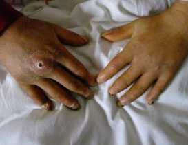
Scleroderma
|
Your Website:Home >>
Scleroderma |
|
|
| Scleroderma laboratory examinations are there? |
| |
Whether localized or system type, involved or affected skin feeling significantly longer time when compared with normal measurement (extended from 5 to 12 times).
Most systems ESR type faster. Some cases of lupus can be found in blood cells. Fluorescent antinuclear antibody positive rate of about 95%, fluorescence karyotype is more common to spot too, also can be seen in visible nucleoli CRST Anticentromere (anticentromere) staining. Immuno diffusion technique measuring anti-Scl-70 antibodies have greater Tete symmetrical diffuse type heterosexual. A fold of skin at the capillary microscopy, revealed that most capillaries blurred significantly reduce leakage and edema, vascular several loops, branching blood vessels dilated and bent, blood flow is slow, many accompanied by bleeding.
X-ray examination: System type patients tend to show: ① widened periodontal ligament; ② esophagus, gastrointestinal peristalsis disappeared, the lower end of a narrow, widened proximal small bowel reduction, expansion of the proximal small intestine, colon bags spherical change; ③ finger bone resorption; ④ two lung markings, or see underestimate cystic change; ⑤ have calcium deposits within the shadow of the soft tissue. |
| |
| Previous:
What is Raynaud‘s phenomenon, Raynaud‘s syndrome?
|
| Next:
Scleroderma typing
|
| [Close] |
|
 |
|